Breast Cancer Screening: Know Your Options
September 4, 2025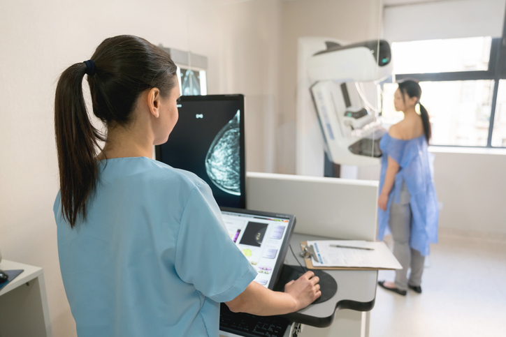
Categories: Cancer Care
Tags: mammography for breast cancer, Breast Cancer, Diagnostic Radiology
Breast cancer screening isn’t one-size-fits-all—it’s a set of tools you can tailor to your risk and preferences. From digital and 3D mammography to ultrasound, breast MRI, and genetic/risk assessments, each option plays a different role in finding cancer earlier, when treatment is most effective.
Here, Allison Heil—Supervisor of Breast Imaging at Riverside Healthcare—shares how these screenings work, who may benefit, and what to discuss with your healthcare provider so you can make a confident, personalized plan that fits your age, health history, and breast density. 
Why Is Mammography So Effective?
Mammograms can find breast cancer when it’s tiny—often years before a lump can be felt. The earliest form, stage 0/DCIS, may appear only as microscopic calcifications that radiologists spot with magnification and can biopsy. Because these changes can take 5–10 years to become palpable, screening catches disease when it’s most treatable. Early detection often means less extensive surgery (e.g. lumpectomy instead of mastectomy) and may reduce the need for additional oncology treatments.
“There are so many factors that go into treating breast cancer,” states Heil. “But, if you find it at its earliest stage, oftentimes you can have success with minimal treatments.”
What Does the Mammogram Entail?
Some women avoid mammograms due to discomfort or result anxiety, but mammography is the only test that can see the earliest calcifications—not visible on ultrasound or MRI—making it the best tool for early detection. The exam is a quick outpatient X-ray of both breasts so the radiologist can compare symmetry. Gentle compression is used to hold the breast still and see tissue clearly. However, Heil urges patients to speak up so the technologist can adjust if anything feels uncomfortable.
“We don't want this to be an exam women don't want to come back to. So, speak up. If the compression is too uncomfortable, let your technologist know. Maybe they can shift your arm, shift the height of the machine,” she assures. “Usually, though, we only need two to three images of each breast. Then, we let you go on your way.”
3D mammography (tomosynthesis) is the standard because it captures multiple thin “slices” of the breast—up to approximately 30, depending on thickness—for a clearer view. It requires a slightly longer exposure, so if a patient can’t comfortably stand, needs to sit, or has limited shoulder mobility, a 2D mammogram is a solid alternative that still produces quality images. In practice, about 98% of patients can tolerate 3D.
Breast Tissue Density Matters
States now require notifying patients of their breast density in both the radiology report and the lay letter after a mammogram. Density is categorized A–D—or described as fatty/fibroglandular, heterogeneously dense, moderately dense, or extremely dense—and can change over time with pregnancy, menopause, and hormone use.
Dense tissue, especially categories C or D, is harder to interpret on mammograms. If you’re C or D, keep up with annual mammography and consider supplemental screening—such as breast MRI or ABUS (automated breast ultrasound). Knowing your density helps you and your provider tailor your screening plan.
“These exams do not replace the mammogram,” notes Heil. “Mammography is the standard screening of choice. But, those two additional tests can help women with dense tissue. It can give the radiologist more information and help them better see through that dense tissue. Also, if you're a high-risk patient, those additional screenings can be beneficial as well.”
What Constitutes a High-Risk Patient?
Riverside uses a risk-assessment software during mammogram visits that combines your history (e.g., age at first period, pregnancies, prior breast surgery, family history) to generate a risk score. Per guidelines, a score over 20% is considered high risk. Radiologists see this score with your images and may recommend supplemental screening—such as breast MRI or ABUS—when appropriate. Many high-risk patients already have known factors, including family history, dense breasts, or prior biopsies showing atypia or papilloma.
Genetic testing is typically pursued in the context of a cancer diagnosis or strong family history and is often coordinated by oncology. But, most people don’t undergo it routinely. For example, if you’re BRCA-positive, you’ll still have annual screening mammograms—with additional high-risk imaging recommended alongside mammography.
One clarifying point Heil makes is that of all breast cancers diagnosed, only 10% are family history-related. “The other ninety percent of women out there who don't have family history are the women getting diagnosed. It's your neighbor or your coworker. They got their mammogram, had a biopsy, and now they're encountering this little detour in life. Just because you're not considered high risk doesn't mean you shouldn't get screened.”
To learn more about mammography and imaging options at Riverside Healthcare, visit myrhc.net/imagingservices.
Related Providers


Related Articles
How a Breast Cancer Support Group Is Healing Together
A grassroots group of breast cancer survivors is proving that healing happens best in community.When Rive...
Men: Get serious about your cancer risk
One of the most important things you can do for your health is talking to your primary care provider (PCP...